About
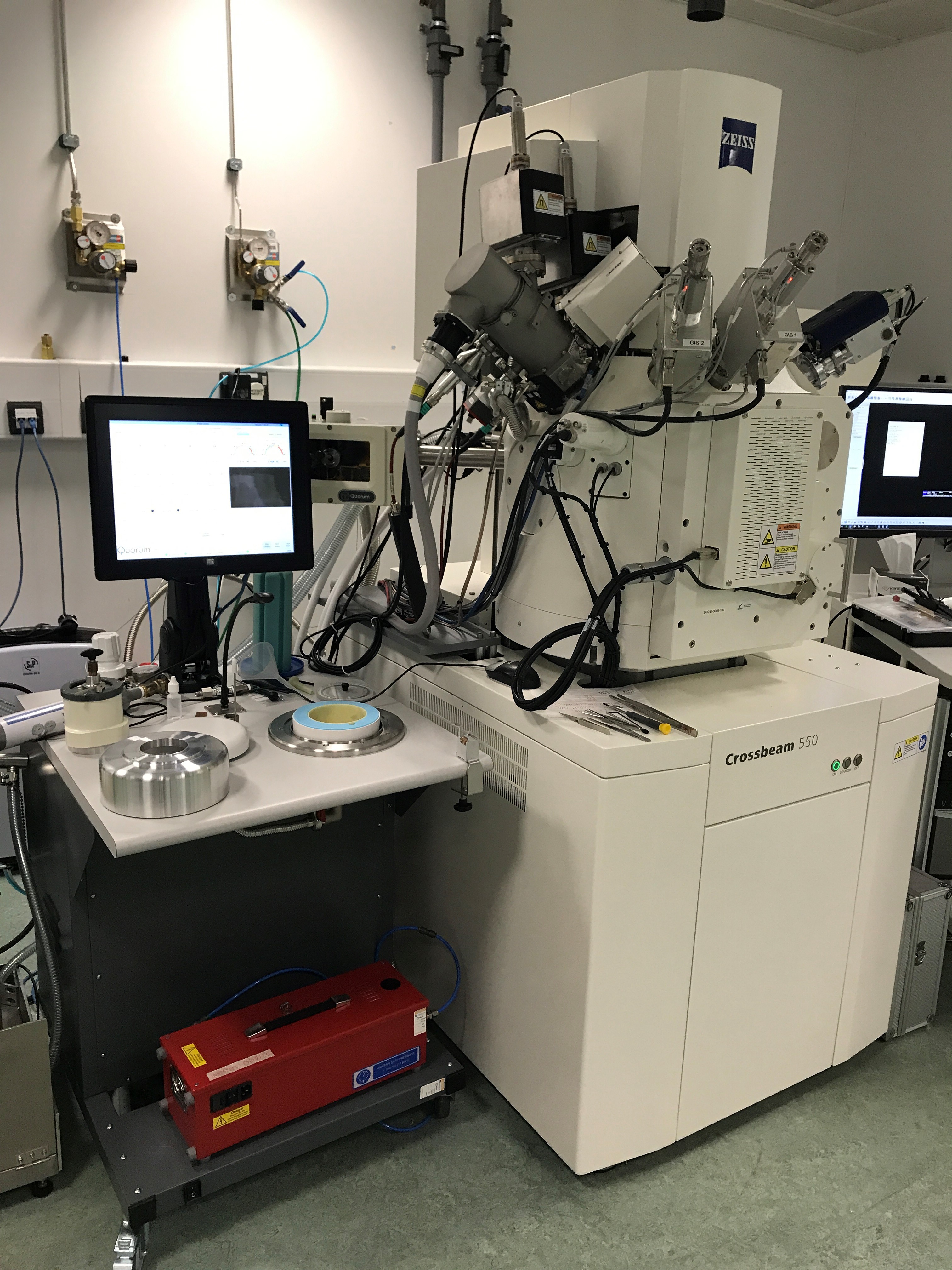 The Crossbeam is a high-resolution FE-SEM equipped with a Gemini II SEM column, a Capella Ga- liquid metal ion (Ga-LMIS) FIB and a Quorum cryo-stage and cryo-prep station with airlock. It is a highly versatile piece of kit which can be used for a range of applications in the life and material sciences, allowing high-resolution SEM imaging at room temperature and in cryogenic conditions. Dual-channel detection is also enabled, allowing surface and material contrast images to be acquired simultaneously.
The FIB beam can be used to mill nanostructures onto material samples, but also to perform slice-and-view destructive 3D imaging and to prepare lamellas for electron tomography experiments. Area refinding between different imaging modalities can be achieved using preinstalled Atlas software, enabling correlative imaging of specimens.
The system also features:
- 6-axis motorised stage
- two Gas Injection Systems, capable of on-stage coating of samples with platinum or carbon;
- plasma-driven platinum cryo-coating device in the Quorum prep station;
- sublimation prep device in Quorum prep station;
- freeze-fracture capabilities in Quorum prep station;
What we provide
- Support and training for room-temperature single-modality or correlative imaging of biological and material specimens, 2D or 3D
- Support and training for cryogenic temperature single-modality or correlative imaging of biological and material specimens, 2D or 3D
- Secure data storage and transfer
- Support and training for data analysis using open source environments such as Fiji, Icy and the MIB-3D Slicer-Blender pipeline
What we do not provide
- Support, training or facilities for metal impregnation and resin embedding of biological samples. The samples will have to be already processed for imaging prior to facility access
Example serial section imaging datasets
Cryogenic serial section imaging of U20S cells with a slice depth of 20 nm.

A 3D render of a manually segmented COVID-19 infected Vero cell acquired using cryogenic serial section imaging. The distribution of COVID virus particles (red) is revealed alongside cell organelles and components such as mitrochondria, nested vesicles, lipid droplets, golgi and the complex membrane. Sample courtesy of the Zhang group, University of Oxford.
Available Detectors
Chamber Everhart Thornley SE2
| Surface contrast, long range, not compatible with slice-and-view imaging
|
InLens SE2
| Surface contrast, short range, compatible with slice-and-view imaging
|
InLens ESB
| Some material contrast, compatible with slice-and-view imaging
|
Pneumatic retractable 4-quadrant solid-state BDS1
| Provides material contrast, not compatible with slice-and view modality
|
Available sample holders
9-stub carousel holder
| Room temperature
|
CorrMic holder for room temperature correlation between Zeiss instruments – best used with tissue sections on slides or EM grids
| Room temperature
|
Flat cryo-holder for bare EM grids
| Cryogenic - Quorum stage
|
Flat cryo-holder for autogrids
| Cryogenic - Quorum stage
|
Flat cryo-holder for bare sapphire disks
| Cryogenic - Quorum stage
|
Flat cryo-holder for superSIL assemblies
| Cryogenic - Quorum stage
|
Flat HPF Planchette cryo-holder
| Cryogenic - Quorum stage
|
Pretilted cryo-holder for bare EM grids
| Cryogenic - Quorum stage
|
Variety of cryo-SEM holders for fibres, foams, gels and solid samples
| Cryogenic - Quorum stage
|
General specifications
Sample temperature range
| 25 - -175°C |
Stage tilt
| -15 - 70°
|
Stage rotation
| 360 continuous (room temperature sample stage only)
|
Column angle
| 54°
|
| Scanning electron gun |
SEM voltage
| 0.02-30 kV
|
SEM probe intensity
| 10 pA - 40 nA (high-resolution mode) or 10 pA -100 nA (high-current mode)
|
SEM magnification
| 12- 2,000,000 x
|
SEM resolution
| 1.6 nm at 1 kV
|
Detector collector bias
| 0 - 400 V
|
ESB grid
| 0 - 1500 V
|
Image format
| 8 or 16 bit tiff, 4:3 format up to 32768 x 24576 pixels
|
Focussed ion beam gun
|
FIB Voltage
| 30 kV (0.5-5 kV in low kV mode)
|
FIB probe intensity
| 1pA – 100 nA
|
FIB magnification
| 300 – 500,000 x
|
FIB resolution
| 3 nm at 30 kV
|
Contact
For further information about the Zeiss Crossbeam at the Octopus facility or to discuss a potential application, please contact Dr. Laura Zanetti-Domingues or Mr. Benji Bateman.