Laser-driven X-ray sources are incredibly valuable for taking images of intricate and fast-moving components. Using them, we can see things in detail far beyond what an ordinary X-ray could – including being able to differentiate between similar materials within a sample that an average X-ray could not. X-ray imaging is a core application of our up-and-coming laser facility, the Extreme Photonics Applications Centre (EPAC).
Recently, a team led by Dr Dan Symes from the CLF in collaboration with the John Adams Institute and Diamond Light Source have been putting this into practice using a range of samples provided by Johnson Matthey (JM), the Warwick Manufacturing Group, the National Composite Centre, and University Hospital Galway.
To get the best out of the experiment, some interesting cross-collaborations have been established. Tomographic imaging is a new area for the high-power laser community, and with that, we have joined the pre-established networks between end-users of advanced X-ray technology and experts from the Collaborative Computational Project in Tomographic Imaging. Within STFC, the project draws on the wealth of experience in image processing and big data storage that is regularly handled by the Scientific Computing Department and Diamond Light Source scientists.
Using the Gemini laser, the team imaged several complex objects such as batteries, car catalytic converters and cancerous tissues. A high quality image of carbon-composit sample was acquired (below) thanks to the spatial coherence of the laser-driven X-ray source in the 10's of keV range, which, when travelling through a sample, results in a phenomenon called phase-contrast enhancement radiography. In weakly absorbing objects, phase imaging is more sensitive to small density changes leading to superior image contrast compared to conventional X-ray scanners.
Since objects such as catalytic converters and carbon fibre materials are made of several slightly different materials, this technique is extremely useful for industry. Oftentimes, objects like these would appear homogeneous on a normal 3D X-ray scan, making it difficult to observe its features. It is also beneficial for biological applications as cancerous cells and normal cells are hard to distinguish with standard radiology.
Dr Dan Symes said about the experiment, “This is all very new technology for applications – it's only really in the last few years that these techniques have been made more robust. So far it has produced some great results."
Another technique that Dr Dan Symes and his team investigated during the experiment was the production of ultrashort pulses of hard X-rays of 100s keV, which can penetrate thick, high-density objects. This is beneficial because they can be used to create high-resolution scans of fast processes within larger samples such as entire engines in motion. This technique is very hard to do, but the CLF plans on building the capabilities to refine this.
This experiment – which has been one in a series over the past few years – is helping the CLF pave the way towards better, faster, and more reliable imaging setups and techniques for EPAC.
Dr Chris Thornton, an EPSRC-UKRI industrial research fellow who worked on the research project, said, "These experiments have shown great promise in offering another imaging dimension to what is available to the X-ray imaging community. Through links with academia and industry, we are learning how best to exploit the unique X-ray source to have the biggest impact on science and industry."
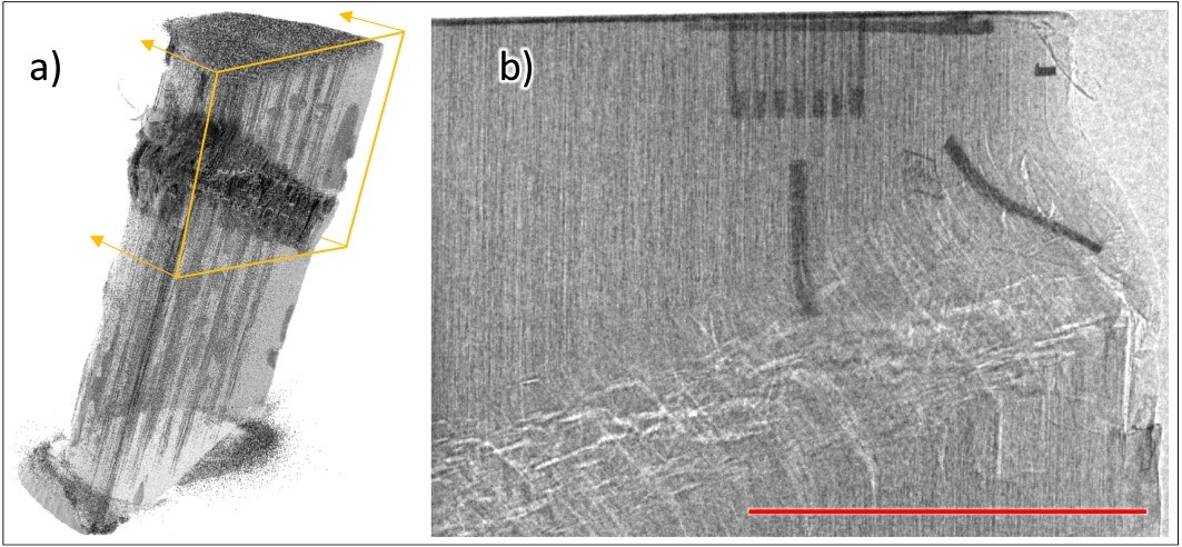
(Kink band failure in a composite cylinder initiated by impact and propagated by compressive end loading. (a) tomographic reconstruction obtained using conventional lab x-ray CT (b) radiograph obtained with the laser-betatron source with carbon fibre tows visible. The red line indicates 1 cm.) Image credit: https://www.sciencedirect.com/science/article/pii/S016890022030766X?via%3Dihub
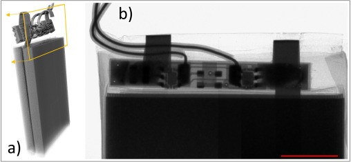
(Tomographic reconstruction of a pouch cell obtained using conventional lab x-ray CT. Manufacturing defects can occur such as delamination of layers, improper electrode attachment, and poor welding in the tab area highlighted (b) Front view radiograph of the tab area obtained with the laser-betatron source. The red line indicates 1 cm.) Image credit: https://www.sciencedirect.com/science/article/pii/S016890022030766X?via%3Dihub
To read the latest paper, click here.
A new paper relating to this experiment will be published soon. The link will be updated on this page.