The minimal photon fluxes (MINFLUX) concept is a novel synthesis of aspects of single molecule localisation microscopy (SMLM) and Stimulated Emission Depletion (STED) microscopy that can achieve far higher spatial and temporal resolution than either, while requiring far fewer fluorescence photons.
Techniques
1. Ultra-high precision 2D and 3D single molecule localisation

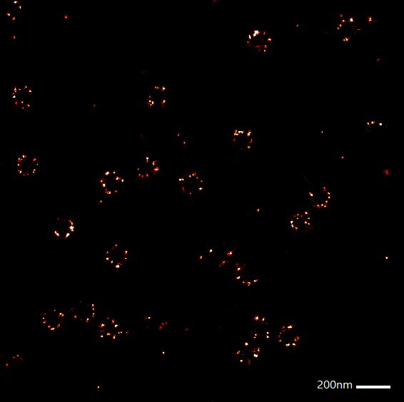
Comparative images of Nup96 complexes labelled with Alexa 647. Left: standard confocal mode, right: 2D Minflux mode.
Nup96, a nuclear pore complex protein labelled with Alexa 647 has been imaged with approximately 1nm x, y resolution. The same sample imaged in 3D yielded x, y precision of 2.4nm and z precision of 1.9nm, clearly resolving Nup96 octamers on either side of the nuclear membrane3. 2 colour MINFLUX can clearly distinguish the centre of the nuclear pore labelled with wheat germ agglutinin from the Nup96 octamer.
MINFLUX localisation nanoscopy has also been applied to PMP70 distribution in peroxisomal lipid membranes, clathrin light chain organisation in membranes and mitochondrial MICOS proteins.
2. Active tracking of molecules with high spatio-temporal resolution
The lipid DPPE labelled with Atto 647N has been tracked in a supported lipid bilayer with localisation precisions <20nm and a mean position sampling rate of 8.5 kHz (∆tmean ≈ 118µs). This is far higher temporal resolution than camera based single molecule tracking with passive localisation. Camera based tracking is restricted to sampling rates of 50-100 Hz and then often suffers from poor localisation precision due to insufficient fluorescence photon flux unless bulky gold particles or quantum dots are used as a probe.
Applications
Biological and medical research.
Specifications
Confocal mode: 488nm and 642nm excitation, GFP/near-IR/far-IR detection channels.
MINFLUX mode: 642nm excitation single probe detection in near- or far-IR channels. 2-probe MINFLUX is possible using 2 probes that can be separated by spectral classification using the two IR channels.
Recommended fluorescent probes for MINFLUX:
Ultra-high precision localisation microscopy
Single probe: Alexa 647
Two probes: Sulfo-Cyanin 5 and CF680
Please note that the required blinking characteristics for successful MINFLUX localisation microscopy is more stringent than for dSTORM. Not all probes that can be used for dSTORM will work with MINFLUX
Ultra-high speed tracking
Atto 647N
Contact
For further information about the Minflux, or to discuss potential applications, please contact Dr. Chris Tynan.