Lasers, as fascinating as they may sound, are complicated to explain, and it is even more complicated to explain their applied use in experiments.
Hand in hand with lasers, spectroscopy plays a huge part in countless CLF experiments, where using different wavelengths of light is the key to identifying minute artefacts.
A symposium and workshop on correlative light, electron and X-ray microscopy was held from the 6th- 10th March this year. Located in STFC's Pickavance Lecture Theatre in Harwell, the event aimed to discuss the developments of these complex fields, and how scientists have been using them to get accurate results in their experiments.
With an aim to inform the community of the latest developments in correlative microscopy, the symposium consisted of lectures from world-leading scientists working in the area of correlative microscopy with light, electrons and X-rays.
During the symposium, a key-note lecture by Martin Saunders from the University of Western Australia discussed the need for many different types of spectroscopy in order to accurately analyse certain artefacts, and how the data from some types of spectroscopy can be misleading if not used in collaboration with other types of spectroscopy. His work on studying the presence of Earth's early life in rocks showed the upsides and pitfalls to all types of spectroscopy – especially when the sample is incredibly rare.
To give an example, Electron microscopy can give you a very accurate image, but only of a very tiny section of the sample. This means that analysing something can take an incredibly long time. Light Microscopy gives a great picture, but is effectively looking at the sample with the human eye, meaning that you can only see the surface of the sample and nothing underneath. You also cannot see if there are hidden substances imbedded in the sample (which a technique like raman spectroscopy could tell you).
By combining light and electron spectroscopy and cutting the sample in a process called CLEM microscopy, you can get a drastically more accurate image, but in the process, the sample is destroyed.
To help explain each type of spectroscopy in a simple way, I drew this illustration after attending part of the symposium.
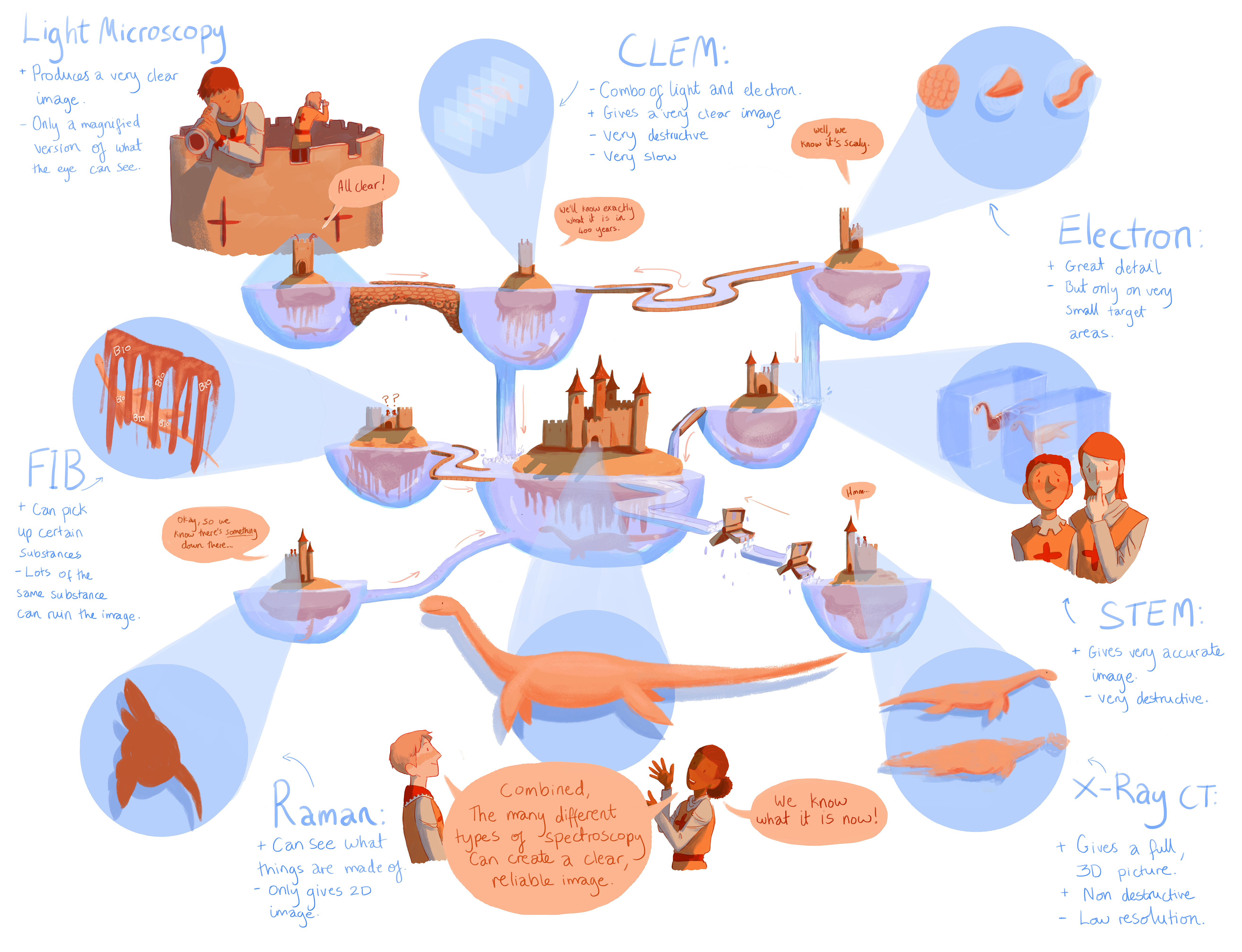

The monster under each moat to symbolises the hidden, difficult to analyse sample, and the people atop the castles are scientists trying to identify the monster.
The CLF hopes to use images like this, aimed at older children, to strengthen the general awareness of analytic processes such as spectroscopy, and to portray them in a dynamic, exciting, and most importantly, understandable way.