Dr Richard Chapman and his team at the CLF in collaboration with Dr Bill Brocklesby, Prof Jeremy Frey and their team from the University of Southampton have used the Artemis facility to image lab-grown neurons originating from mice, revealing extraordinary detail compared to traditional light microscopic images. The extra detail available means that the technique has potential applications in medicine, particularly in studying neurodegenerative diseases such as Alzheimer's disease.
From Baksh et al., Sci. Adv. 2020; 6 : eaaz3025. This work is licensed under CC BY (https://creativecommons.org/licenses/by/4.0/).
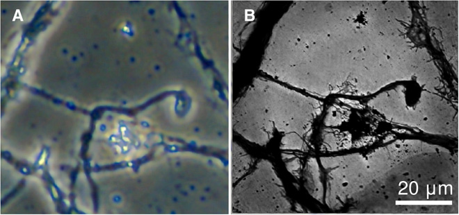
This study has built on the techniques Dr Brocklesby's team developed for EUV lensless microscopy. It consisted of producing coherent extreme ultraviolet (EUV) light from an ultrafast laser to collect scattered light from the samples. Multiple scatter patterns from different sample regions are processed by a computer algorithm to give the highly detailed images. Comparison of traditional light microscope images (figure 2(a) above) and the EUV images (Figure 2(b) shows that much finer details are visible using EUV.
EUV imaging has been performed at large, expensive facilities such as synchrotrons and free electron lasers. However, using laser sources makes this a compact and lower-cost setup that can mean this technology is more accessible. The recent upgrade to the Artemis facility means that it will be able to push further into the EUV region, allowing for even higher resolution images. Combining these images with techniques available in the Octopus facility at the CLF allow for steps towards developing correlative imaging tools.
The results of this study combined with the factors highlighted above means that EUV techniques on Artemis carry the potential to observe the structure of neurons in great detail, enabling scientists to identify morphological changes related to disease. Following these results, the Artemis team is working towards being able to offer regular access to this technique in the future.
To read the paper, click here: https://advances.sciencemag.org/content/6/18/eaaz3025


