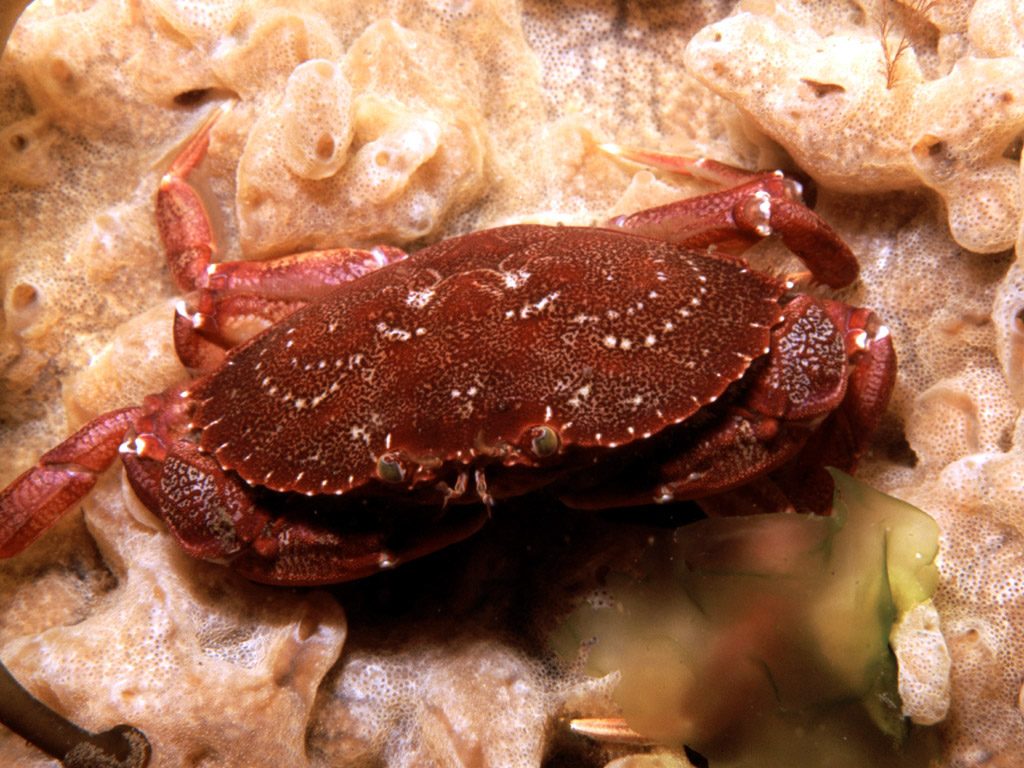
Crustacean shell disease is a condition that damages the outer skeleton of crabs and leads to significant impacts on their health. The initial shell damage, which allows bacteria and microbes resulting in shell disease to invade, is largely triggered by fighting, predation, and even cannibalism between crabs.
A recent study led by Prof Sigrun Lange at the University of Westminster, UK, in collaboration with the University of Iceland and STFC's Central Laser Facility (CLF), has applied high-resolution microscopy techniques for the first time to identify extracellular vesicles (EVs) as new biomarkers in rock crab shell disease. EVs are tiny, membrane-bound particles released by cells to facilitate cell-to-cell communication.
To date, EVs have mainly been used as biomarkers for monitoring human disease, but the study's findings provide a strong foundation for further development of this technique for marine and environmental applications.
Infectious and invasive
First discovered in Icelandic waters in 2006, the Atlantic rock crab (Cancer irroratus) has already outcompeted native crab species through predation, habitat competition or indirect ecosystem cascades.
The invasive status of C. irroratus provided the ecological motivation for this study, as changes in their health and distribution in an environment where they have few natural predators may disproportionately affect local marine species.
Although Atlantic rock crabs could emerge as a valuable seafood resource to Iceland, with their naturally fleshy legs and growing numbers in warming Icelandic waters, they are fundamentally an invasive species that poses a threat to native biodiversity.
The cell's stealthy messengers
This new collaborative study reports that crab shell disease causes changes to the EVs circulating in the animals' blood-like fluid (known in crustaceans as haemolymph). EVs are microscopic packages constantly released by cells, carrying biological messages in the form of protein, mRNA and DNA as part of cell-cell communication.
Up to now, EVs have been extensively researched for use as biomarkers in human diseases, including cancer and neurodegenerative diseases but this new research suggests that EVs may also be useful and just as important as disease biomarkers in marine species.
Identification of crab haemolymph EV signatures
In this study, haemolymph was collected from male and female Atlantic rock crabs, which were captured at five sampling stations in Hvalfjörður, a 35-kilometre-long fjord in southwest Iceland. The research team then isolated haemolymph EVs and compared their signatures in male and female crabs with and without visible shell disease.
The team used high-resolution dSTORM microscopy at the CLF's Octopus imaging cluster, for imaging EV markers, and assessed the protein content of the EVs. dSTORM imaging revealed changes in vesicle types in both sexes, and the number of EVs was increased in the infected male crabs, which may be related to the infection. The varying EV protein cargo between sexes may indicate that responses to shell disease in males and females occur via slightly different physiological pathways, but the reasons for these differences remain unclear and will require further study.
Protein analysis of the EV content revealed 34 different proteins, some of which changed with disease and differed between the sexes. Some of the proteins found only in the EVs of infected animals are associated with immune defence, cellular communication, protein production and DNA repair.
The findings suggest that detecting these EV biomarkers will help scientists assess immune response pathways and support monitoring of diseases and environmental stress in crabs, or other commercially or ecologically important crustaceans.
While this study was conducted as a proof of principle, it provides a foundation for developing future diagnostic tests following larger studies.
Making the invisible visible at LSF–Octopus
With EVs at only 40 to 500 nanometres in size, access to dSTORM super-resolution microscopy at the LSF's Octopus imaging cluster was essential to be able to label and quantify these minuscule crab vesicles.
CLF researchers Dr Sarah Needham and Dr Ben Davis contributed crucial imaging resources, methodology, data curation and analysis, allowing researchers to:
- visualise individual extracellular vesicles
- distinguish different types of vesicles using fluorescent (glowing) labels
- identify infection-related differences that varied between male and female crabs
- combine these highly detailed imaging results with measurements from other techniques (such as nanoparticle tracking and electron microscopy)
Future disease epidemic monitoring
This study was the first to use dSTORM to visualise EVs in crustaceans, establishing a new imaging approach for investigating biomarkers during infection.
Lead author Prof Lange, Head of the Pathobiology and Extracellular Vesicles Research Group at the University of Westminster, is a pioneer in EV research across the tree of life, including marine species. She said:
“The development of novel techniques for EV research is still at early stages in animal research, where the potential in aquaculture and farming is huge, as well as for wild biology research. Working together in international and interdisciplinary teams, bringing together a wide range of technologies and expertise, we are able to push the boundaries in global research, relevant for the future of our planet."
With further research, EV biomarkers could potentially be used to study disease epidemics in marine species globally. As ocean temperatures and pollution levels continue to change, monitoring marine health and disease with novel sensitive tools will be critical for understanding the complex aquatic ecosystems, which are fundamental to fisheries and global food security.
Visit the Comparative Immunology Reports paper “Extracellular vesicle signatures are modified in haemolymph of Atlantic rock crab (Cancer irroratus) infected with shell disease – a Study from Icelandic waters" here.