At the Central Laser Facility, we collaborate closely with many world-class institutions to carry out cutting-edge medical research. Our principal current projects are summarised below.
Supra-molecular rules in the HER signalling network in cells and tissues
Dr Marisa Martin-Fernandez, with Prof Peter Parker, Prof Tony Ng, Prof George Santis, Dr Simon Ameer-Beg, Prof Ton Coolen (all King’s College London), Prof Mike Hobson (University of Cambridge), Dr Martyn Winn (Daresbury Laboratory) & Prof Mark Sansom (University of Oxford).
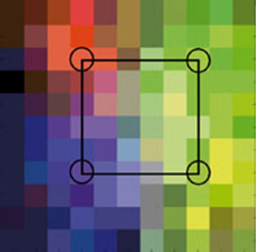
We are investigating the patterns of structural conformation, association, activation and signal transduction for the four members of the HER family (HER1-4) and their correlation with plasma membrane proximal receptor traffic and downstream signalling using 2D cultures of cell lines. We are deriving models of ligand-induced behaviour and establishing how they are influenced by feed-back loops and internal and external perturbations, e.g. ErbB mutations (seen in human subjects), receptor tyrosine kinase (RTK) inhibitors (RTKIs) and function-blocking antibodies. These models will be tested on primary airway epithelial cells and lung tissues and adapted iteratively to create a framework of response prediction for the network. The work uses a cross-disciplinary portfolio of advanced experimental techniques, computational simulation, systems modelling and analysis techniques, many of which have been developed for this specific project but are widely applicable to our user programs. These include multicolour single-molecule microscopes and Bayesian image feature detection algorithms. Ultimately, we will develop methodologies, probes and/or algorithms based on the models that we have derived that are suited to the interrogation of fixed or fresh human tissues to determine the robustness of the models and the predictability of the network in a real world setting.
Inhibitor-induced HER2-HER3 heterodimerisation promotes proliferation through a novel dimer interface
Claus, J et al
ELIFE (2018)
Oligomerization of the Epidermal Growth Factor Receptor Organizes Kinase-Active Dimers into Competent Signaling Platforms
Needham, S et al
BIOPHYSICAL JOURNAL 112 (3): 26a-27a (2017)
A tale of the epidermal growth factor receptor: The quest for structural resolution on cells
Tynan, C et al
METHODS (95): 86-93 (2016)
EGFR oligomerization organizes kinase-active dimers into competent signalling platforms
Needham, S et al
NATURE COMMUNICATIONS (7): 13307 (2016)
Effect of Phosphorylation on EGFR Dimer Stability Probed by Single-Molecule Dynamics and FRET/FLIM
Coban, O et al
BIOPHYSICAL JOURNAL 108 (5): 1013-1026 (2015)
Structure-function relationships and supramolecular organization of the EGFR (epidermal growth factor receptor) on the cell surface
Needham, S et al
BIOCHEMICAL SOCIETY TRANSACTIONS 42 (1): 114-119 (2014)
Novel Platform Technology for the Next Generation of Biomedical Sensors
 Prof Pavel Matousek, with Prof Duncan Graham (University of Strathclyde) & Prof Nicholas Stone (University of Exeter)
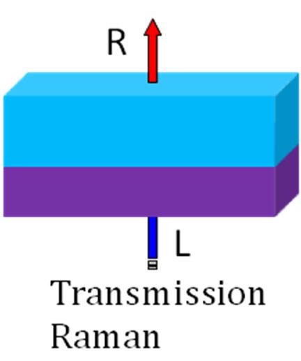
The project aims to develop the optical sensors for disease diagnosis by monitoring bio-analytes at extremely low concentrations. This is achieved by combining Spatially Offset Raman Spectroscopy (SORS) with traditional SERS (surface enhanced Raman spectroscopy), in a so called SESORS modality. The team has succeeded in demonstrating a unique deep imaging capability of SESORS by measuring signals from within a 50 mm-thick slab of biological tissue. This record breaking penetration depth is an order of magnitude higher than demonstrated previously with conventional SERS and paves the way for the next generation of optical biosensors.
Noninvasive Determination of Depth in Transmission Raman Spectroscopy in Turbid Media Based on Sample Differential Transmittance
Gardner, B; Stone, N; Matousek, P;
ANALYTICAL CHEMISTRY: 89 (18) 9730-9733 (2017)
Studying the distribution of deep Raman spectroscopy signals using liquid tissue phantoms with varying optical properties
Vardaki, M; Gardner, B; Stone, N; Matousek, P
ANALYST: (15) (2015)
Recent advances in the development of Raman spectroscopy for deep non-invasive medical diagnosis
Matousek, P; Stone, N
JOURNAL OF BIOPHOTONICS: (6); 1 (2012)
Surface enhanced spatially offset Raman spectroscopic (SESORS) imaging – the next dimension
Stone, N; Kerssens, MM; Lloyd, GR; Faulds, K; Graham, D; Matousek, P
CHEMICAL SCIENCE 2: 776-780 (2011)
Prospects of Deep Raman Spectroscopy for Noninvasive Detection of Conjugated Surface Enhanced Resonance Raman Scattering Nanoparticles Buried within 25 mm of Mammalian Tissue
Stone, N; Faulds, K; Graham, D; Matousek, P
ANALYTICAL CHEMISTRY 82: 3969–3973 (2010)
Development of Novel Non-invasive Breast Cancer Diagnosis
A new project, led by Professor Nick Stone from the University of Exeter and funded by EPSRC, will use the emerging field of nanotheranostics, combining therapy and diagnosis, to identify and treat life-threatening diseases in a single, effective non-surgical procedure.
Nanotheranostics derives from the term “theranostics" which refers to a form of diagnostic testing that allows a patient to receive selective targeted therapy. Cancer nanotheranostics aims to combine imaging and cancer therapy using nanotechnology. By engineering nanomaterials to  interact with light and cancer cells at a nanometre scale level, the effectiveness and specificity of cancer therapy could be substantially increased
The new research project will use light to identify early changes within the body and deliver treatment or monitor its progression, without resorting to invasive tissue removing techniques. Harnessing the principle of nanotechnology, the group will develop a new approach for assembling tiny gold nanoparticle clusters within the body that will allow selective targeting of diseased cells, avoiding healthy tissue. One of the key technologies behind the project, Spatially Offset Raman Spectroscopy (SORS) originates from the Central Laser Facility and was jointly developed for cancer diagnostic applications in partnership with the University of Exeter.
High sensitivity non-invasive detection of calcifications deep inside biological tissue using Transmission Raman Spectroscopy
Ghita, A; Matousek, P; Stone, N
J BIOPHOTONICS Jan;11(1) (2018)
Development of deep subsurface Raman spectroscopy for medical diagnosis and disease monitoring
Matousek, P; Stone, N
CHEMICAL SOCIETY REVIEWS 45, 1794-1802 (2016)
Non-invasive chemically specific measurement of subsurface temperature in biological tissues using surface-enhanced spatially offset Raman spectroscopy
Gardner, B; Matousek, P; Stone, N
FARADAY DISCUSSIONS 187, 329-339(2016)
Recent advances in the development of Raman spectroscopy for deep non-invasive medical diagnosis
Matousek, P; Stone, N
J BIOPHOTONICS, Jan;6(1): 7-19 (2013)
Towards a safe non-invasive method for evaluating the carbonate substitution levels of hydroxyapatite (HAP) in micro-calcifications found in breast tissue
Kerssens, MM; Matousek, P; Rogers, K; Stone, N
ANALYST 135: 3156-3161 (2010)
Advanced Transmission Raman Spectroscopy: A Promising Tool for Breast Disease Diagnosis
Stone, N; Matousek, P
CANCER RESEARCH 68: 4424-4430 (2008)
Prospects for the Diagnosis of Breast Cancer by Non-Invasive Probing of Calcifications using Transmission Raman Spectroscopy
Matousek, P; Stone, N
JOURNAL OF BIOMEDICAL OPTICS 12: 024008 (2007)
Development of Novel Noninvasive Bone Disease Diagnosis
 Prof Pavel Matousek, Prof Anthony Parker & Dr Kevin Buckley, with Prof Allen Goodship (University College London) in collaboration with the Royal National Orthopaedic Hospital (Stanmore)
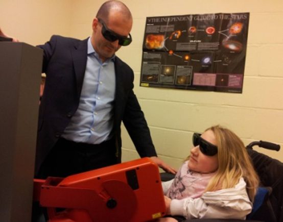
The CLF is developing a new method to detect early signs of diseases such as brittle bone disease and osteoarthritis. The study centres on Spatially Offset Raman Spectroscopy (SORS) developed within the CLF and the work is being lead by Professor Allen Goodship. At present we are taking a series of measurements from volunteers with the long term aim of developing a specialist medical device. This is very early work but our goal is to eventually validate a new method to enable better preventive treatments to be taken at an early stage of disease development. The rationale is that diagnosing disease earlier leads to more effective treatment. This new study, funded by the EPSRC (grant number EP/H002693/1 (link opens in a new window)), paves the way for future patient clinical trials to validate potential wider applications such as screening for osteoporosis and connective tissue disorders. A major advantage of using Raman spectroscopy is that it gives a molecular finger-print of the material making up the bone, measuring both the inorganic (mineral) and organic (collagen/protein) phase; in contrast, the main method used at present uses X-rays to characterise the bone but this only able to study the mineral phase.


