When medical procedures (like radiotherapy) use radiation to damage 'dangerous' cells (for example cancer cells), this is not a helpful process as it can greatly hinder the treatments effectiveness. Professor Stanley Botchway at Octopus has been investigating how and why cells are triggered to repair their DNA after radiation damage. This will hopefully help improve the effectiveness of medical treatments that rely on radiation and chemicals permanently damaging (and sometimes terminating) cells.
The research conducted at the CLF-Octopus, and at CLF high power lasers Vulcan and Gemini, in collaboration with UK- Health Security Agency and Oxford University, uses standard x-rays, laser generated exotic particles, alpha particles, and direct pulsed laser in the near infra-red (NIR) to mimic ionising radiation.
The use of NIR light and a multiphoton process in particular means the cell DNA damage only occurs in a femtolitre volume. This highly localised deposition of energy means the cells are not overwhelmed during the DNA damage and they can be studied for longer.
NIR light is not significantly absorbed by cells and tissues whilst if UV is used, this will damage most organelles and proteins inside the cell, making any outcome more complex to understand. Furthermore, since the DNA damage occurs under a microscope, the repair process can be visualised in real time and under conditions that are ideal for the cells, such as at 37 degrees Celsius.
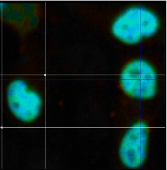
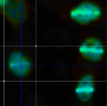
The images on the right hand side contain cells showing cell nuclei before (top image) and after (bottom image) laser induced damage. The scale bar is 10 mm.


