Applications of the techniques
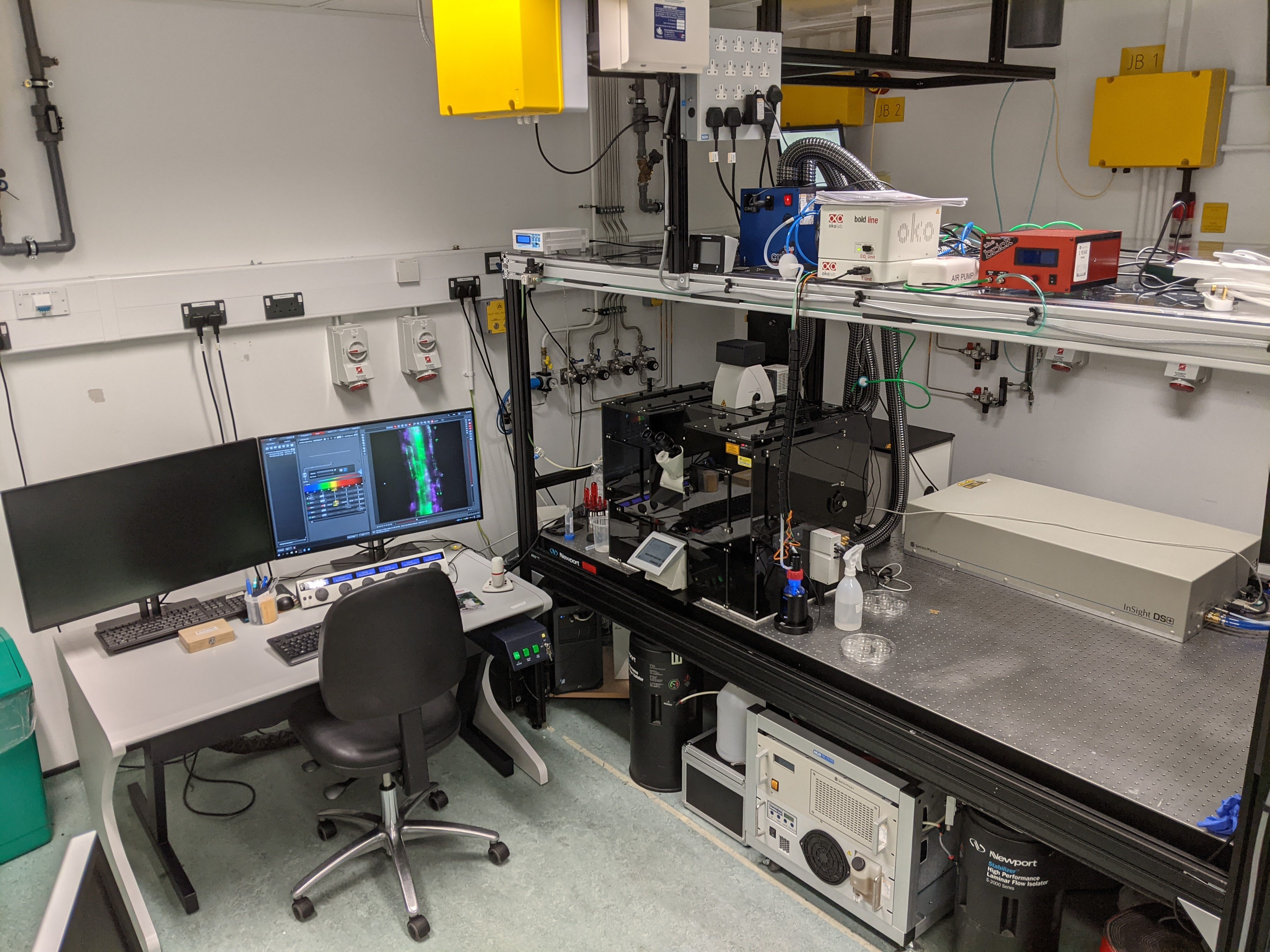
At Octopus, we have two lightsheet fluorescence microscopes capable of imaging a range of samples which are live, fixed or cleared, the Leica SP8 Digital Light Sheet (DLS) and the 3i Cleared Tissue Light Sheet (CTLS). The Leica SP8 DLS is also capable of confocal, multiphoton and lifetime imaging in an inverted setup (the objectives are below the sample) which is most well-suited to slides/coverslips and dishes with glass bases.
The Leica SP8 DLS is used for samples that are thinner than 1 mm, or samples where you only need a 1 mm field of view/slice (i.e. part of an organism). Spheroids and organoids are perfect for this system, along with small transparent organisms (e.g. zebrafish) where only a small area needs to be imaged (e.g. section of the brain). Additionally, cells growing inside of a 3D gel are suitable for this system. We have also used it for more innovative applications such as cells flowing through tubing/nozzles during bioprinting.
The Leica SP8 DLS is suitable for live, fixed and cleared samples (however you are limited to 1-2 micrometre lateral resolution for cleared samples). There is also an environmental chamber to control CO2 levels and temperature.
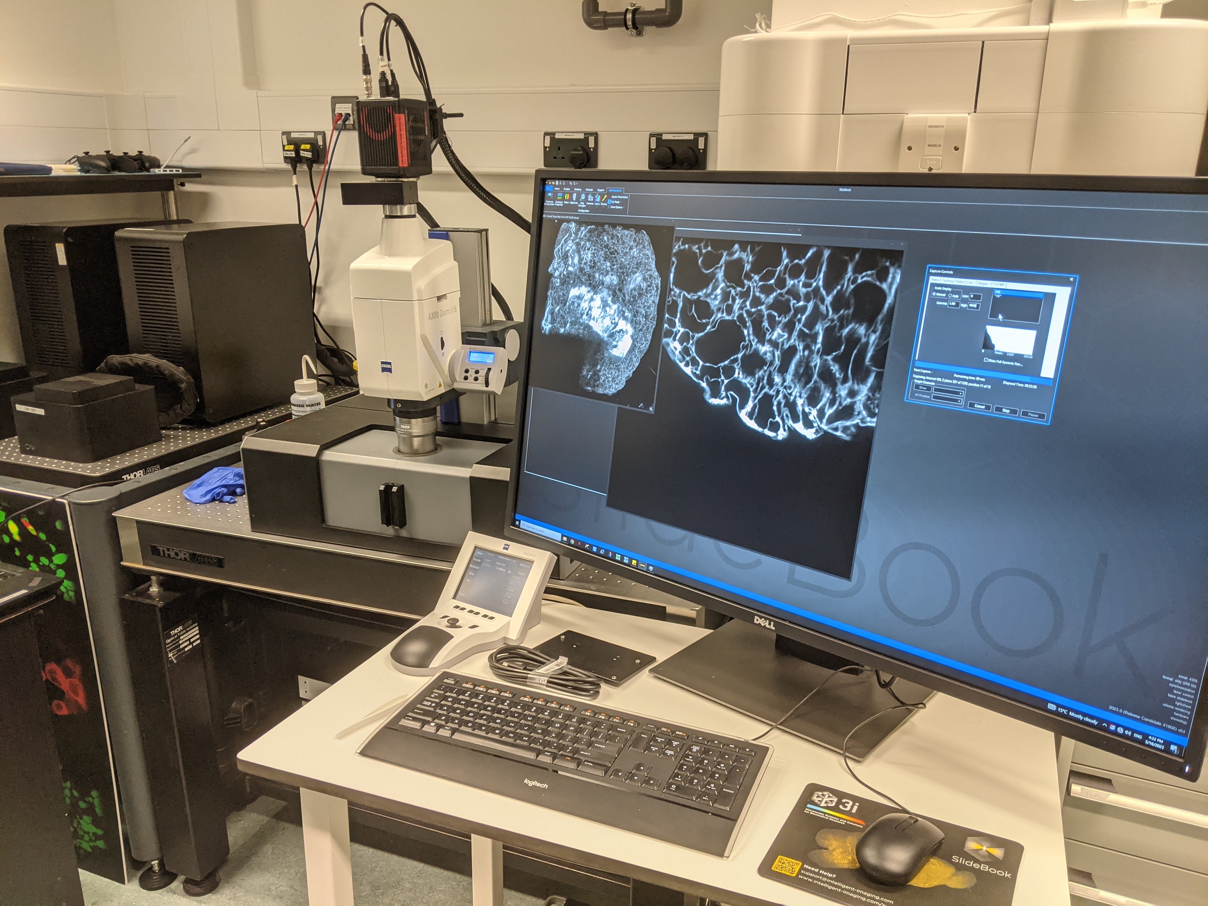
The 3i CTLS is suitable for live or fixed clear samples, whether that is artificial (through clearing) or natural. For live samples, they must be viable at room temperature as there is no environmental control. It can handle relatively large samples up to the size of about 20 x 50 x 18 mm, or multiple samples in a high throughput organisation. This is suitable for imaging cells embedded in gels, cleared tissue samples from human (biopsies) and animals, including whole organs (kidney, liver, eyes etc.) or even whole organisms (neonatal mice).
If you have an application that you think is suited to one of our microscopes, get in touch, we'd be happy to advise you on whether the instrument can be used for your needs. For further information please contact Dr. Robert Lees.
Technical specifications
Below are the specifications for the microscopes, the resolution is not listed as it is highly variable depending on the combination of illumination and detection objectives and also is linked to the size of the image you can get. Typically you will be getting 1 um lateral resolution and <5 um axial resolution for a 500 micrometre+ field of view, however much higher resolution is possible on the Leica SP8 and there is some ability on the 3i CTLS to get higher resolution if you sacrifice total sample size imaged.
Leica SP8 DLS Specifications
Lasers: White light laser (80-10MHz) 470-680 nm free choice 405 nm CW OPO laser for MP 80 MHz 680-1300 nm free choice Detectors: Hamamatsu ORCA Flash 4.0 V2 sCMOS Camera,
2 internal photon counting HyD, 2 internal PMTs, 2 external cooled HyD, 2 external PMTs, trans-PMT
Objectives: Light sheet detection: 5x W, 10x W, 25x W Confocal and multiphoton: 63X W, 63X Oil, 40X W, 25X IR W, 10X, 5X
Filters:
Light sheet: 500-620 nm + 655-750 nm dual band, 504-545 nm, 575-635 nm, 405/488/561/633 nm quad-band Confocal and multiphoton: internal detectors from 380-800 nm, external detectors fixed at 500-550 nm, 435-485 nm, 565-605 nm, 467-499 nm, 430-450 nm
|
3i CTLS Specifications
Lasers: Solid state diode lasers combined using a LaserStack: 405, 445, 488, 514, 561, 594, 640 nm
Detectors: Hamamatsu ORCA Flash 4.0 V3 sCMOS Camera
Objectives: Light sheet detection: 1.0x/0.25 Air, 1.5x/0.37 Air, 2.3x/0.57 Air
Light sheet illumination: 5x/0.14 Air, 10x/0.28 Air
Filters:
Light sheet: quad-band 446/523/600/677-25 nm, 505-545 nm, 573-637 nm, 580-654 nm, 663-733 nm
|


