Scientists at CLF have collaborated with researchers at Polintecnico di Milano to characterise porcine tissue. The results show a large variation in the optical properties of the tissues. The data can be used to construct artificial structures mimicking specific diseases exploiting the wide assortment in optical properties and guide the development of optical disease diagnostic techniques.
The work done here serves as a major advancement in the field of optical characterisation of animal tissue as it synthesises much of the previous work done on the subject in a more comprehensive and consistent way. Previous optical characterisations of living tissue have been found to be inadequate for several reasons: The data was inconsistent due to the source tissue coming from a variety of different animals which were often only reported at narrow wavelengths and was limited in the type of organ tissue it came from. Furthermore, these results were obtained using different experimental and theoretical approaches which led to inconsistent data sets.
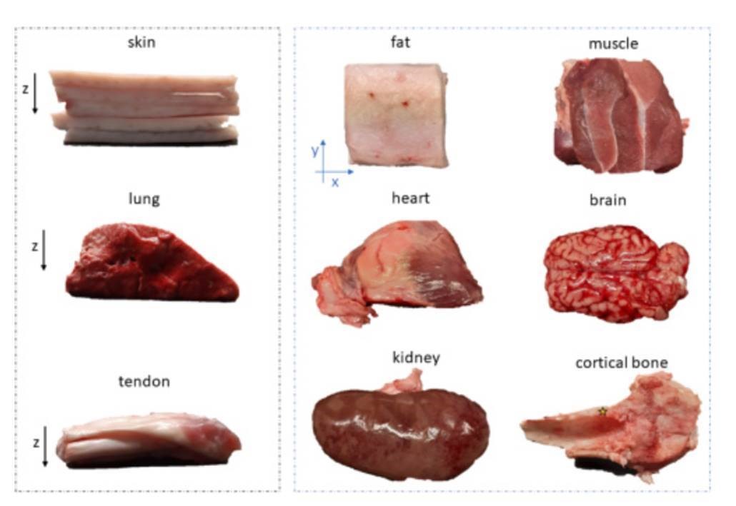
The study conducted by the team was much more comprehensive, taking into account a greater range of parameters and more tissue variety to create a more general and useful model for future biomedical research. Porcine tissue was specifically chosen as it shares many of the key characteristics of human tissue that are relevant in therapeutic testing. Some of the selection of porcine tissues for optical characterisation included skin, brain, bone, tendon, adipose, muscle, kidney, heart and lung tissue.
To achieve this, the team characterised these porcine ex vivo tissues using a technique called TD-DOS (Time-Domain Diffuse Optical Spectroscopy). This approach deals with light propagation in diffusive media and enables unambiguous dis-entanglement of absorption and reduced scattering coefficients of tissues and can also be implemented over a broad range in the near-infrared spectral region. For this purpose, the scientists used a broadband TD-DOS system at Politecnico di Milano.
The results of this study are broadly consistent with the sporadic data available in previous works, and will help to create accurate light transport models for predicting the light propagation and for the optimization of experimental optical parameters of tissue samples. Having access to the optical properties of the same animal species allows one to create a model that mimics heterogeneous structure by combining different tissue types.
With a more comprehensive and consistent characterisation of the various different types of tissue, it is hoped that more accurate assessment of disease models can be made which would be valuable resource in biomedical research.
Funded by the Horizon 2020 Framework Programme (654148).
Link to the original paper here: https://www.osapublishing.org/boe/fulltext.cfm?uri=boe-11-3-1697&id=427973
Image Ref: Photos of all 9 porcine ex-vivo samples. The yellow star in the cortical bone panel indicates the analyzed point. https://www.osapublishing.org/boe/fulltext.cfm?uri=boe-11-3-1697&id=427973#figanchor2


