High energy density physics (HEDP) experiments constitute research in fields ranging from condensed matter to meteorite impacts, from material failure mechanisms to the behaviour of planetary cores. States such as these, with high temperatures and pressures, are accessible in the laboratory through dynamic compression experiments – a shock wave is driven into a material, with this particular experiment, by a pulsed laser.
For more than a decade at Imperial College London, Dr Stuart Mangles and Dr Daniel Eakins (who proposed the research), along with a research team have been developing a new type of compact accelerator called a laser wakefield accelerator which has been used in this experiment. It has the capability to make ultrafast flashes of X-rays that can 'freeze' the motion of rapidly changing systems.
“The X-rays produced by the laser wakefield accelerator at Gemini are great for taking snapshots of shocks as they fly through some material because they are very short flashes of X-rays."
Dr Stuart Mangles (Imperial College London)
Furthermore, the use of betatron radiation – ultrashort pulsed, hard, bright X-rays – for radiography is an enticing foray into the imaging of rapidly evolving phenomena. As it stands presently, traditional surface based measurements are proving inadequate as the subsurface interactions cannot be diagnosed directly. The result being, measurements of shock properties at the surface are potentially affected by interactions below the surface which are not being observed.
The Gemini laser at the Central Laser Facility, with its two beamlines, proved invaluable as the Gemini South beam was used in the production of the betatron radiation and the Gemini North beam was used to produce the shock wave in the target. The delay between the target being hit and the betatron probe was able to be varied continuously between 0 and 12 nanoseconds allowing for snapshots to be taken of the shock wave. One benefit proposed by betatron radiation is that rapidly evolving phenomenon can be imaged without the need for larger, more expensive, light synchrotron sources.
The footprint of the experiment detailed, had the target placed 0.12m from the source and the camera was 3.70m from the source. When considering the scale of X-ray imaging beamlines from synchrotrons, which are typically around 100m long, the benefit of using an experimental setup this small is immense. It allows ultrafast, hard X-ray probing experiments to be performed at university scale facilities and at a fraction of the cost! Alternatively, a betatron imaging beamline could be implemented at large scale HEDP facilities, again lowering the cost. Further still, due to the high flux of betatron X-rays, standard detectors were sufficient to allow single pulse imaging.
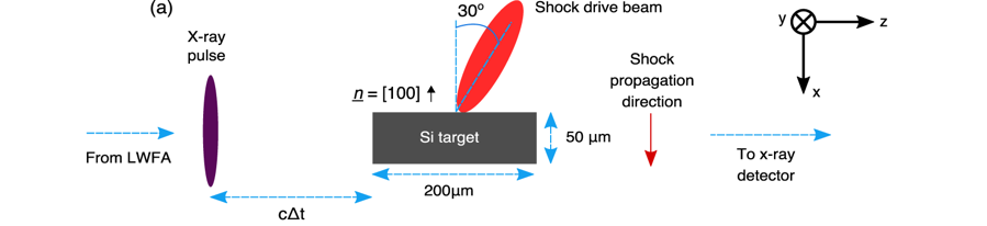

Figure 1: a) Top down view of the shock target interaction point. Image credit, Imperial College London.
As confirmation of this proof of principle experiment, the shock velocity was measured from the position of the shock fronts – a technique permitted by the clarity of the images as seen in Figure 2. The research team were also able to measure the compressed density from the measured opacity of the target and the known properties of the X-ray source. The density of the silicon undergoing compressions was simulated and the experimental findings were in agreement with the simulation as well as data obtained from other experiments. The experimental result for the shock velocity was found to be 6.2+-0.4 km/s and from the simulation 6.7 km/s.

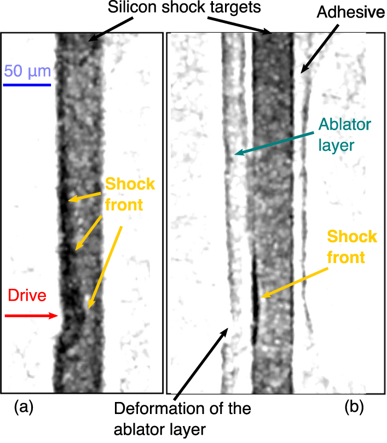
Figure 2: a) X-Ray image of shocked silicon target at 5.2ns after the start of the interaction. b) X-ray image of shock wave in silicon with an ablator layer on the drive surface at 6.5ns. Image credit, Imperial College London.
Overall, the outcome shows that the spatial resolution of the image is comparable to results of similar experiments at light synchrotron sources. However, faster processes could be imaged as the temporal resolution of betatron radiation is below 100 femtoseconds. The future of compact betatron radiation imaging will see the technique enter the fray of high energy density physics experiments, due to the versatility of the source, allowing it to be coupled to a variety of drivers.
Dr Stuart Mangles had this to say about the direction the research is heading in the future:
“Now that we've successfully demonstrated imaging of things that move on a nanosecond timescale we are looking at ways to use the ultrashort X-ray duration to probe systems that evolve even more rapidly: techniques such as X-ray absorption spectroscopy or diffraction could reveal the rapid processes that occur when we heat matter to extremely high temperatures and pressures that are usually only found in the centre of giant planets and stars."
Dr Stuart Mangles (Imperial College London)
The research was supported by the Science and Technologies Facilities Council, Engineering and Physical Sciences Research Council and the European Research Council.
The full publication is available to view in Scientific Reports
For further information about the research, please contact Dr Jonathan Wood (jonathan.wood08@imperial.ac.uk).
Images used are under open access.
Creative Common's license is available to view here.


