As you are reading this sentence, the iron-rich protein in your body, haemoglobin, is carrying oxygen to your body's organs and tissues and transporting unwanted carbon dioxide back to your lungs. Human haemoglobin (Hb) is composed of two ɑ and two β subunits, where each subunit can interact with oxygen to mediate its transport.
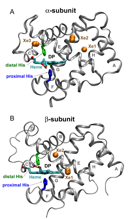
Despite years of research, there isn't a universal agreement on the protein's function. Researchers are still trying to map out how alpha and beta subunits binding to oxygen affect the individual behaviour of these building blocks. To highlight how these conformational changes affect the individual ɑ and β subunits when interacting with oxygen, researchers, Sergei V. Lepeshkevich, Marina V. Parkhats, Syargey N. Gilevich and Boris M. Dzhagarov from the National Academy of Sciences of Belarus collaborated with CLF scientist, Igor V. Sazanovich, on a study published in the Royal Society of Chemistry journal.
For the experiment, researchers created a model species that mimic haemoglobin's behaviour. Next, at the Ultra facility, the individual alpha and beta subunits were subjected to time-resolved absorption spectroscopy, so researchers could look at conformational changes within milliseconds since reactions in a biological system take place at such a speedy rate.
The study revealed that alpha and beta subunits in haemoglobin behave very differently from each other when bound to oxygen and that the hybrid species that were made to mimic haemoglobin are extremely good models. From a biological standpoint, this research is extremely important because it bridges our gap in understanding how the individual subunits function within the whole of haemoglobin and how oxygen transport takes place.
Moreover, the research also highlights the potential of laser science in biological research. As researchers were firing laser pulses at the haemoglobin species, the heme group – which resides in the centre of the haemoglobin – releases oxygen, which leads to a colour change that researchers can follow. As a result, the Ultra laser system was functioning akin to a sophisticated oximeter. Just like an oximeter detects oxygen level by measuring the red light transmission through the tissue, our laser system can do that more accurately at an advanced resolution. Therefore lasers, such as the one present in Ultra, provide a novel insights into complex biological systems, such as that in haemoglobin.
Futher Information:
Full paper can be found at DOI:https://doi.org/10.1039/D1SC00712B


