This large-scale multidisciplinary work received funding from many different national institutes, including the Wellcome Trust, the NIHR Great Ormond Street Hospital Biomedical Research Centre, Kidney Research UK, and the NHS Foundation Trust, and was led by Dr Daniyal Jafree alongside Prof David Long (both University College London (UCL)).
The current kidney blueprint
Within our bodies, interstitial fluids comprise approximately 11 litres of blood-like watery fluid fill the spaces between cells and tissues. Kidney lymphatic networks are a system of specialised vessels responsible for maintaining overall volume and composition of the interstitial fluid. By draining proteins, immune cells, and interstitial fluid from kidney tissues and returning them to the blood, the kidney lymphatic vessels play a crucial role in maintaining overall bodily fluid balance. Despite this, the role of these vessels in building a healthy kidney is poorly understood.
Kidney lymphatic network vessels (known as glomeruli) are formed during prenatal development; these help determine the kidney's filtering capacity throughout life, with a lower number associated with greater susceptibility to kidney disease. By studying kidney assembly during development, scientists can gain a greater understanding of kidney structure and diseases.
Beyond the naked eye
This study focused on two specific diseases in the kidneys of genetically modified mice. When imaged under the microscope, the clinicians could see by eye that one diseased vessel network looked more complicated than the other, with the vessels filling the space differently. When trying to quantify these differences using conventional methods, for example by measuring average vessel length, thickness and number of branches, the vessel networks between diseases were indistinguishable: despite obvious differences in their organisation, the individual vessels that made up the network had similar structures. Alternative approaches were therefore needed to quantify the differences between these vessel networks in the kidney, to better understand how fluid flows in and out of these tissues.
Nature's mathematical fingerprint
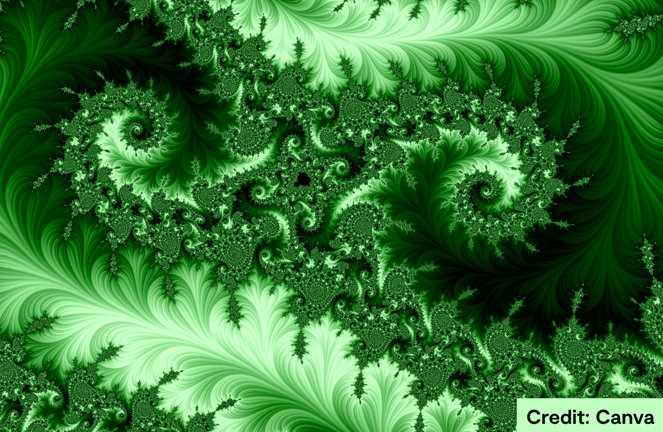
Fractals are a mathematical set or geometric shape found wherever patterns repeat across different scales. Fractal patterns, which become increasingly complex as you zoom in, are commonly found in nature, including in fern fronds, snowflakes, river networks, lightning bolts and coastlines. While natural structures often show imperfect versions of this perfect mathematical behaviour, fractals are an extremely useful model in measuring and comparing the properties of nature's intricate, branching networks.
Fractals (from the Latin word fractus, meaning broken) were developed as a field of mathematics by Benoît Mandelbrot in the 1970s. Mandelbrot however, credits some of this development to earlier work by Lewis Fry Richardson, a British scientist who was trying to measure the length of the British coastline and noticed that the smaller the ruler he used, the longer the total length of the coastline he measured.
For instance, a coastline could be measured using a 1km ruler giving a defined perimeter. If that same coastline were measured in finer detail using a 30cm ruler, it would appear significantly longer, as more jagged and irregular details would be captured. This 'coastline paradox' represents a counterintuitive phenomenon whereby a coastline can occupy a finite area while appearing to have an infinite and ever-increasing perimeter when measured at progressively smaller scales.
Researchers employ this concept of how similar an object looks when measured at different scales (self-similarity) to complex biological structures. While fractals have several mathematical properties, the team used the 'fractal dimension' to capture the overall complexity of the developing kidney lymphatic network as a single number.
Dr Hannah Mitchell at Queen's University Belfast explains that: “Two vascular networks can contain similar lengths and volumes of vessels but be organised in fundamentally different ways. Fractal dimension allowed us to look beyond those conventional averages and measure how the lymphatic network branches and fills space across multiple scales."
In this Cell Reports study, the CLF's Octopus imaging cluster at the Lasers for Science Facility helped exploit fractal and topological data analysis to maximise information extraction from images, discovering structural differences in vessel networks previously undetected by traditional methods.
Decoding the kidney's branching blueprint
Fractal analysis helped support one of the study's central conclusions: that the kidney receives lymphatic cells from two developmental sources, one of which is completely unique to the kidney. Disrupting the DNA coding for this unique source of cells via genetic knockout impairs lymphatic network complexity, making the network much simpler.
This reduced complexity showed that the relatively small population of kidney-specific lymphatic cells (around 15% of the total) plays a pivotal role in normal lymphatic assembly and optimal glomeruli development. This unique source (called the Osr1 mesoderm) provides a developmental explanation for why kidney lymphatics look different from lymphatics in other organs.
A picture paints a thousand words
The kidneys were imaged by confocal microscopy, segmented into binary representations of lymphatic networks, highlighting vessels and tissue components only. Traditional methods of quantifying kidney lymphatic vessels necessitate imposing metrics onto the image or drawing comparisons between images and datasets. By solely utilising the image, fractal analysis is not vulnerable to external measurements and is therefore far better suited to the study of complex interacting structural features, such as patterns of nerve cell loss or the flow properties in a lymphatic network.
Dr Benjamin Davis from the CLF highlights this advantage of fractal analysis compared with traditional metrics, stating: ''All the information is in the image!"
Credit: Cell Reports paper. Visit the Cell Reports website ''Osr1-expressing mesoderm contributes to lymphatic vessel assembly and complexity in the mammalian kidney" here. Imaged by Dr Daniyal Jafree and Prof David Long.
Critical collaborations
Uncovering this hidden change depended on collaboration between clinicians at Great Ormond Street Hospital and UCL, mathematicians at Queen's University Belfast, and data scientists from the CLF's Octopus group. This unique combination of expertise and instruments brought advanced imaging together with mathematics, software and data science, and helped researchers turn highly complex biological images into discoveries relevant to human health.
Insight to impact
With therapeutic manipulation as the ultimate goal, the data team was able to overcome the limitations of standard vascular metrics by turning imaging data into insight, revealing disease-associated alterations in vascular organisation and complexity. By uncovering a previously unrecognised source of kidney lymphatic cells, scientists gained a better understanding of kidney lymphatics as a determinant of kidney disease progression, transplant outcomes, and potentially future regenerative therapies.
Academic and industrial users can apply to access the Octopus facility through its Facility Access Panel. Click here to check for open access calls.
Visit the Cell Reports website to read the paper on ''Osr1-expressing mesoderm contributes to lymphatic vessel assembly and complexity in the mammalian kidney".
The UCL research team worked in affiliation with the UCL Great Ormond Street Institute of Child Health, the UCL Centre for Kidney and Bladder Health and the Wellcome Sanger Institute.
This research was funded by the following diverse contributors:
- Wellcome Trust
- Kidney Research UK
- Rosetrees Trust
- The Foulkes Foundation
- UCL Bogue Research Fellowships
- Medical Research Council
- National Institute for Health and Care Research (NIHR)
- The Royal Society
- The Lister Institute for Preventive Medicine
- NIHR Newcastle Biomedical Research Centre
- NIHR Biomedical Research Centre at Great Ormond Street Hospital for Children NHS Foundation Trust
- University College London