For more information please contact: Dr. Chris Tynan.
Instrument and technique summary
Technique and applications
| Specifications
|
Technique:
Total Internal Reflection (SM-TIRF) Microscopy Wide-field multidimensional fluorescence imaging where signal is limited to 100 nm layer adjacent to coverslip.
Applications:
Biological and medical research, material science, chemistry
| Lasers: 405, 488, 532, 561, 642 nm or white-light excitation. Three-colour simultaneous imaging is standard, but up to six channels
are possible with prior consultation.
Detectors: Andor iXon EMCCDs. Objectives: 100x TIRF oil immersion. Extra information: Directed photo-bleaching and photo-activation available with the new
iLas2 system. Excitation and emission polarisation are resolvable
with prior consultation.
|
Single molecule fluorescence sensitivity is achieved by using Total Internal Reflection Fluorescence (TIRF) or High Inclination illumination to restrict background fluorescence, using extremely sensitive cooled EMCCDs and judicious sample preparation to minimise non-specific probe binding and molecular crowding in the sample.
Single molecule fluorescence imaging allows a fluorescently labelled target to be studied without ensemble averaging of its behaviour. It can be used with a range of commercially available fluorescent dyes, commonly used fluorescent proteins or quantum dots, and applied to a wide range of sample types. It can be combined with directed photo-bleaching or photo-activation of fluorescence and it is particularly useful for investigating biological systems. OCTOPUS single molecule microscopes have been heavily utilised for studying the behaviour of membrane proteins at the surface of adherent cultured cells, antibody captured suspension cultured cells and the membrane of plant cells.
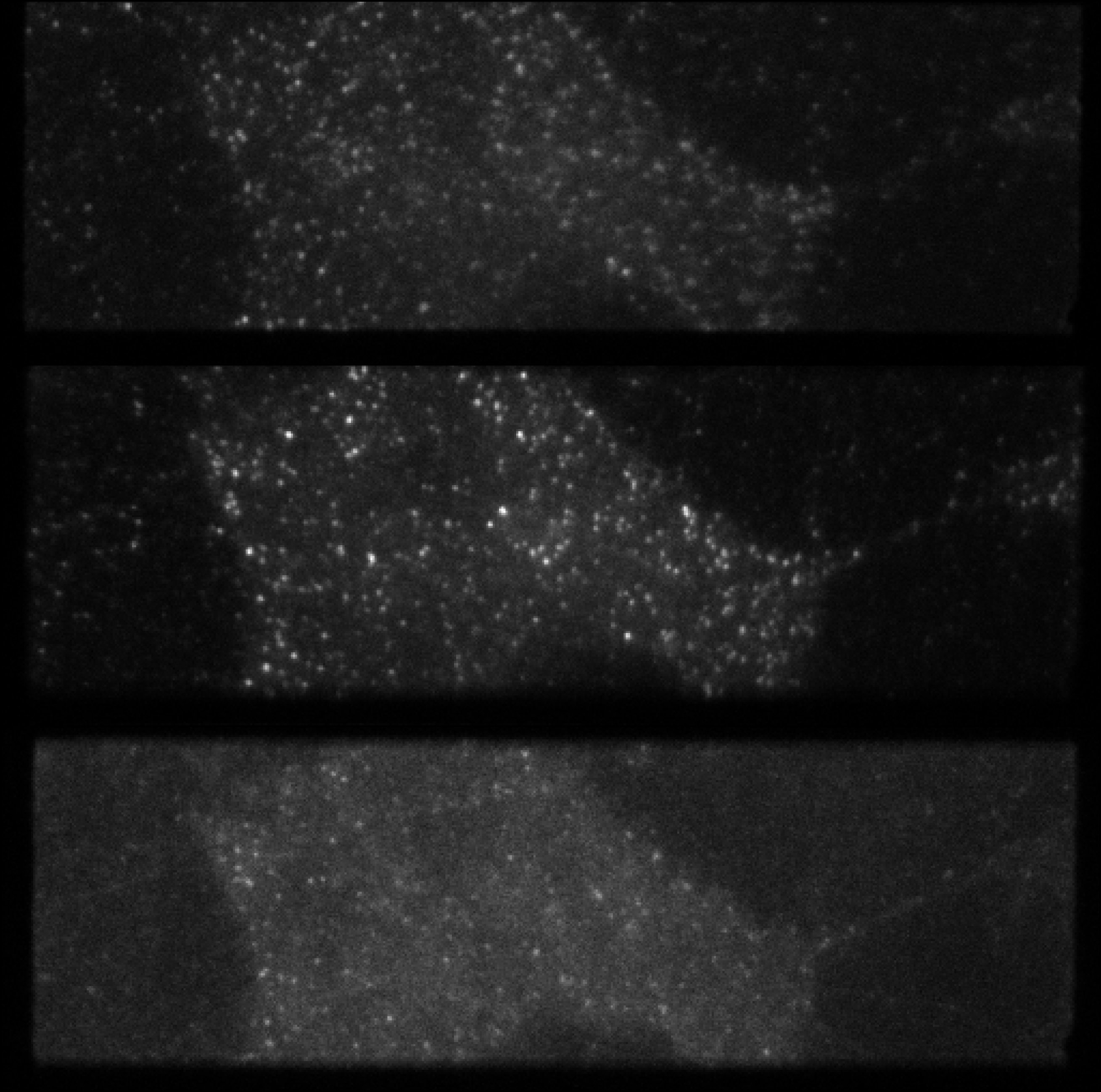
Multi-channel TIRF image of HeLa cells
Technical specification
Imaging modes
Epi-fluorescence, Total Internal Reflection Fluorescence (TIRF)
Illumination/fluorescence excitation sources
405nm, 488nm, 561nm, 640nm Continuous Wave excitation
Available channels and detectors
1 - 6 channels split by wavelength onto Andor iXon cooled EMCCDs. Maximum frame rate is 50 Hz but may need to be reduced to obtain a higher signal from single fluorescent molecules (faster frame rates are possible by reducing the effective chip size).
Techniques available
Multicolour TIRF-microscopy, Single Particle Tracking (SPT), Fluorescence Localisation Imaging with Photo-bleaching (FLImP), Single Molecule-FRET (SM-FRET), SPT after directed photo-bleaching and photo-activation.
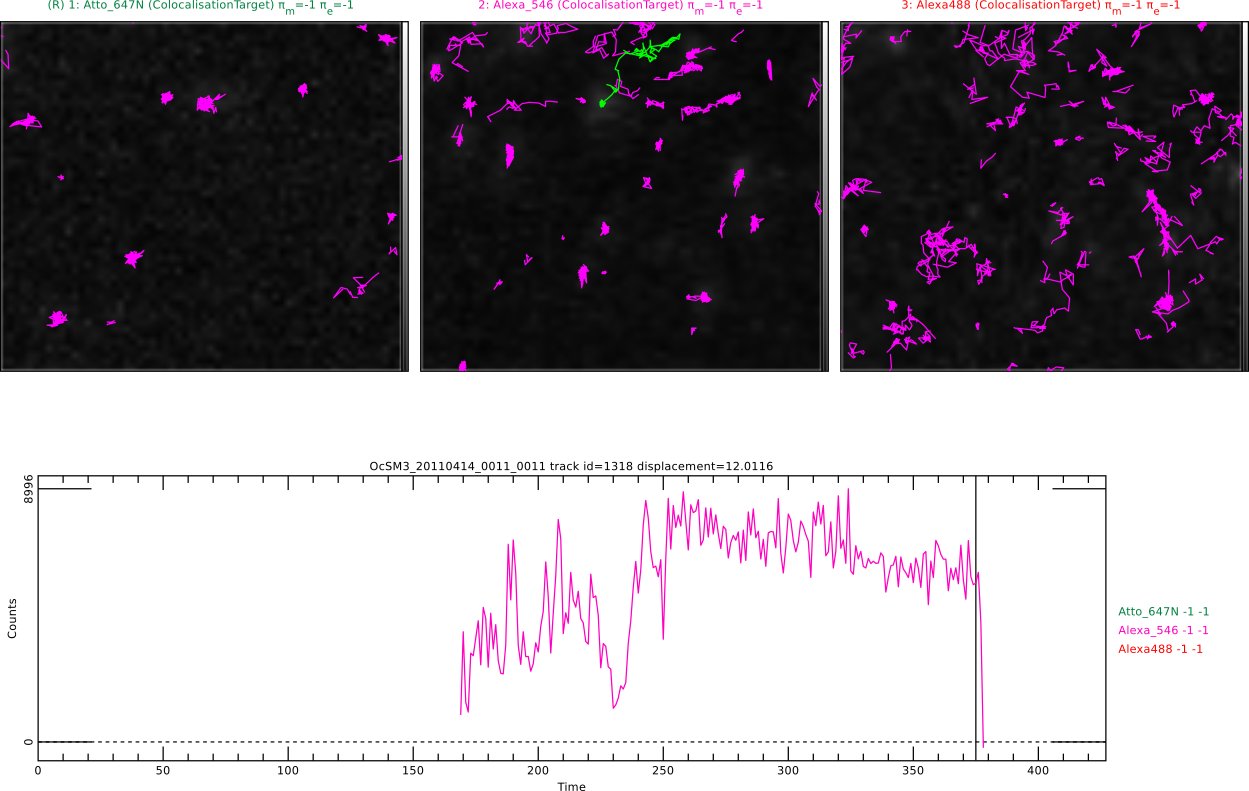
Single molecule tracks reveal co-localisation of fluorescently labelled features
Sample environment
Samples can be created in glass bottom dishes, 4 or 8 well glass bottom slides or slide mounted coverslips. Biological samples can be live (for SPT) or chemically fixed (for FRET and FLImP). Temperature control is available in the range 18-40°C.
Case studies


