Technique and applications
| Specifications
|
Technique:
Becker and Hickl SPC830, SPC150 and SPC230
Wide-field employing Andor ICCD, possible combination with TIRF Applications:
Chemistry solution phase, biomaterial, advanced functional material, catalysis
| Lasers: 405, 488, 543, 642nm laser wavelengths and white light Objectives: 10x, 40x air, 20x NA 0.75, 63x oil immersion, 60x water immersion NA 1.2, 60x air ELWD, 100x oil immersion NA 1.49 Extra information: Fluorescence FLIM 25 ps-50 ns
Phosphorescence PLIM 100 ns-ms
Multi-time 25 ps – 100 ms combined FLIM-PLIM
Multi-colour 2-16 channels
Multi-wavelength excitation
Multi-spectral imaging.
|
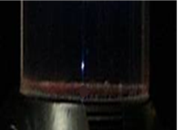 Two-photon (750 nm) excitation of 7-Oh-CCA
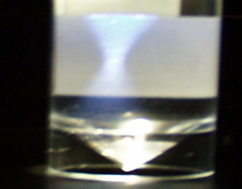 One-photon (375 nm) excitation of 7-Oh-CCA
Laser scanning confocal microscopy offers an advantage over wide-field fluorescence microscopy when using thick samples such as tissues together with high fluorophore labelling densities. This is simply due to interference from emitted photons from everywhere in the sample reaching the detector, leading to blurring of the final image. Confocal uses an aperture (or pin-hole) before the detector to eliminate the out-of-focus plane photons. However the use of the pin-hole and high scattering crossing of the excitation light (~500 nm) means very high photo toxic light levels are used in confocal imaging. Furthermore this leads to reduced photon collection efficiency. Considering the signal to noise ratio (SNR) is proportional to the square root of the detected photon, a pin-hole significantly affects SNR in a negative way.
Two photon or multiphoton excitation (first proposed by Maria Göppert-Mayer) and confocal generally uses near Infra-red to excite the sample and it does not need a pin-hole. This is because the two-photon excitation arises from a simultaneous absorption of two photons in a single quantitised non-linear event within 10E-18 seconds.
Multiphoton imaging provides optical sectioning capabilities to depths of greater than 1mm at diffraction limit resolution. Compared to confocal, multiphoton imaging allows deeper imaging into thick tissue, making it the technique of choice for thicker tissue samples, such as tumour or spheroids as well is intravital imaging in small animal models. Octopus routinely uses multiphoton microscopy for both monolayer cell imaging (reduced photo toxicity) and thick samples such as tissue mimics, biopsy and tumour surrogates, e.g., spheroids.
Microscope full specifications
| Instrument | Nikon EC1, EC2, Becker and Hickl DCS120 |
| Laser wavelengths | Ti:Sapphire laser, Mira F900, 720-980nm, Coherent APE OPO 550-640nm |
| Compatible objective lenses | 10x, 40x air, 20x NA 0.75, 63x oil immersion, 60x water immersion NA 1.2, 60x air ELWD, 100x oil immersion NA 1.49 |
| Compatible dyes | Any |
| Compatible sample mounts | Cover slip on microscope slide or glass bottom Petri dish |
| Configuration | Inverted microscope |
| Temperature control | Environmental chamber or heated stage insert, 5-50 °C. gases include N2, CO2, O2, Ar or any mixture |
| Lateral resolution | ~350 nm (depends on wavelength) |
| Axial resolution | ~800 nm (depends on wavelength) |
| Temporal resolution | From 25 ps (combined with FLIM) |
Applications
- Mammalian and plant cell biology
- Protein-protein interactions
- Time-lapse imaging of cellular processes
- Biological research
- Medical research
- Drug discovery
- Material science
- Geological samples
Case Study
The mammalian target of rapamycin complex 1 (mTORC1) protein interactions (PDF - 59kB - link opens in a new window)


